Handmade Student Models

Physical Disease Models

Handmade physical models built to visualize pathogen structure and connect microbiology to real-world public health in schools.

About These Models

These physical models were hand-built from scratch as part of my ISM project to visually represent the structure of pathogens responsible for school-acquired infections. Building each model deepened my understanding of how these diseases work and how to prevent them.

1

Structure of a Bacterial Cell

Physical 3D model & labeled poster board — clay, paper, and mixed craft materials
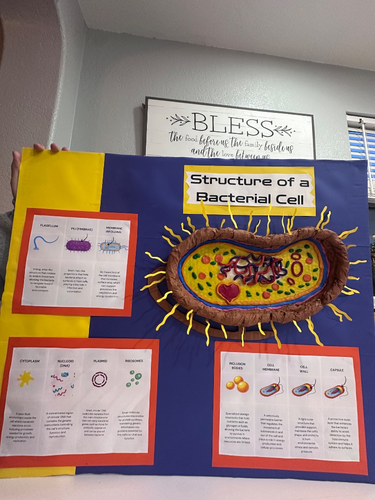
Bacterial cell structure poster board showing labeled cross-section with flagellum, pili, cytoplasm, nucleoid DNA, plasmid, ribosomes, cell membrane, cell wall, and capsule
Click to enlarge

Labeled Components

  • Flagellum
  • Pili (Fimbriae)
  • Capsule
  • Cell Wall & Cell Membrane
  • Cytoplasm
  • Nucleoid (DNA)
  • Plasmid
  • Ribosomes

Public Health Connection

Pertussis (Whooping Cough)

Pertussis Connection

Bordetella pertussis uses fimbriae and pili to colonize the respiratory tract. The plasmid-like elements in this model represent the mobile genetic elements that help bacteria adapt and resist antibiotics over time.

Why This Matters for Treatment

Understanding bacterial structure explains how antibiotics work: cell-wall inhibitors (penicillin, amoxicillin) target the cell wall; macrolides (azithromycin) target the ribosomes. This is why antibiotic choice depends on the type of bacteria causing the infection.

2

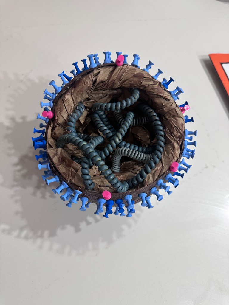
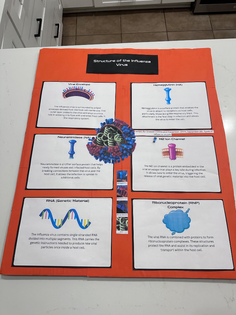
Structure of the Influenza Virus

Physical 3D model & labeled poster board — foam, craft materials, and printed diagrams

Top-down 3D view of handmade influenza virus model showing hemagglutinin surface spikes and internal RNA coils
Click to enlarge
Influenza virus structure poster board showing labeled viral envelope, hemagglutinin, neuraminidase, M2 ion channel, RNA segments, and ribonucleoprotein complex
Click to enlarge

Labeled Components

  • Viral Envelope
  • Hemagglutinin (HA)
  • Neuraminidase (NA)
  • M2 Ion Channel
  • RNA (Genetic Material)
  • Ribonucleoprotein (RNP) Complex
Influenza (Flu)

Why HA & NA Matter in Schools

Hemagglutinin (HA) allows the influenza virus to bind to cells in the respiratory tract, while Neuraminidase (NA) helps newly formed viruses escape the host cell. Because children in schools are in close contact, these surface proteins allow the virus to spread rapidly through classrooms and shared air.

How Vaccines Target This Model

Annual influenza vaccines are formulated to produce antibodies against the HA and NA proteins shown in this model. When HA changes through antigenic drift, the vaccine must be updated — which is why a new flu vaccine is developed every year.

Antiviral Drugs & This Model

Oseltamivir (Tamiflu) and zanamivir (Relenza) are neuraminidase inhibitors — they block the NA protein on this model, preventing the virus from budding out of infected cells. These antivirals are most effective when taken within 48 hours of symptom onset.

How These Models Support the ISM Project

Deepens Scientific Understanding

Building these models required me to research each pathogen's structure in depth — understanding the function of every labeled component and how it contributes to infection, immunity, or treatment.

Supports Audience Differentiation

The models serve as visual aids that communicate pathogen structure at different levels — from a simple visual for elementary students to a detailed structural reference for high school biology classes.

Connects Biology to Prevention

Each component of these models maps directly to a public health action — from the viral surface proteins that vaccines target, to the bacterial cell wall that antibiotics destroy. Biology explains prevention.

Connects to School Populations

The diseases modeled here — influenza and pertussis — are among the most common causes of school absences. Understanding their structure helps students and teachers recognize why prevention strategies like vaccination and handwashing actually work.